Contents
Introduction
Specimens of skin constitute a significant proportion of the workload of many histopathology departments. They are often the first types of specimens that both pathologists and biomedical scientists encounter when they learn the process of cut up. Skin specimens are well suited for the start of this learning process because they are generally simple, illustrate some basic principles and require little variation in technique. Conversely, these same features also limit their usefulness for gaining deeper skill in cut up, especially with regard to correlating observation with block selection, mainly because skin specimens tend to be embedded whole. Nevertheless, the observation and description of a skin specimen mayplay a vital role in the final report because the macroscopic features may alert the pathologist to dual pathology or variation in a single lesion that requires microscopic correlation.
Anatomy
Although often overlooked in the list of structures that are classed as organs, the skin can be claimed actually to be the largest organ in the body. It may be thought of in three layers, the epidermis, dermis and subcutaneous mature adipose tissue.
Epidermis
The epidermis is formed by keratinising stratified squamous epithelium. Melanocytes are located at the base of the epidermis, at a ratio of around one melanocyte per ten keratinocytes. Variations in skin pigmentation are due to differences in the degree of melanin production by the melanocytes rather than in the number of melanocytes.
Dermis
The dermis is located beneath the epidermis and is formed of collagenous connective tissue with admixed elastic fibres. Downward projections of the epidermis, known as rete ridges, interdigitate with the superficial dermis. The dermis between the rete ridges is called the papillary dermis. The dermis deep to this is the reticular dermis. A complex network of cell adhesion molecules and basement membranes allow the epidermis to remain tightly adherent to the dermis.
Various skin adnexal structures are situated in the dermis
|
Hair follicles
|
The density of hair follicles varies greatly depending on the part of the body. Hair follicles are associated with a small muscle, composed of smooth muscle, known as the arrector pili, which causes the hair to stand upright. The arrector pili muscles are responsible for the effect of 'goose bumps'. In evolutionary terms, these muscles were useful for increasing heat insulation in fur-covered animals because the erect hairs trapped a layer of still air which act as additional insulation.
|
|
Sebaceous glands
|
Sebaceous glands are associated with a hair follicle and open into the follicle towards the base of the follicle. They secrete the oily substance sebum which lubricates the hair shaft. The combination of a hair follicle and its associated sebaceous gland is referred to as the pilosebaceous unit
|
|
Eccrine glands
|
Eccrine glands produce sweat and are found in skin from most parts of the body
|
|
Apocrine glands
|
Apocrine also produce sweat but are restricted to the axillary, nipple and genital regions. It is postulated that they are the source of pheremone synthesis. The association of apocrine glands and hair in the axillae and genital region has been suggested to reflect the role of the hair in trapping the apocrine secretions and therefore the pheremonal smell.
|
The dermis will also features blood vessels and nerve fibres.
Subcutaneous Mature Adipose Tissue
The subcutaneous mature adipose tissue is a layer of fat that is located beneath the dermis. It assists in thermal insulation.
|
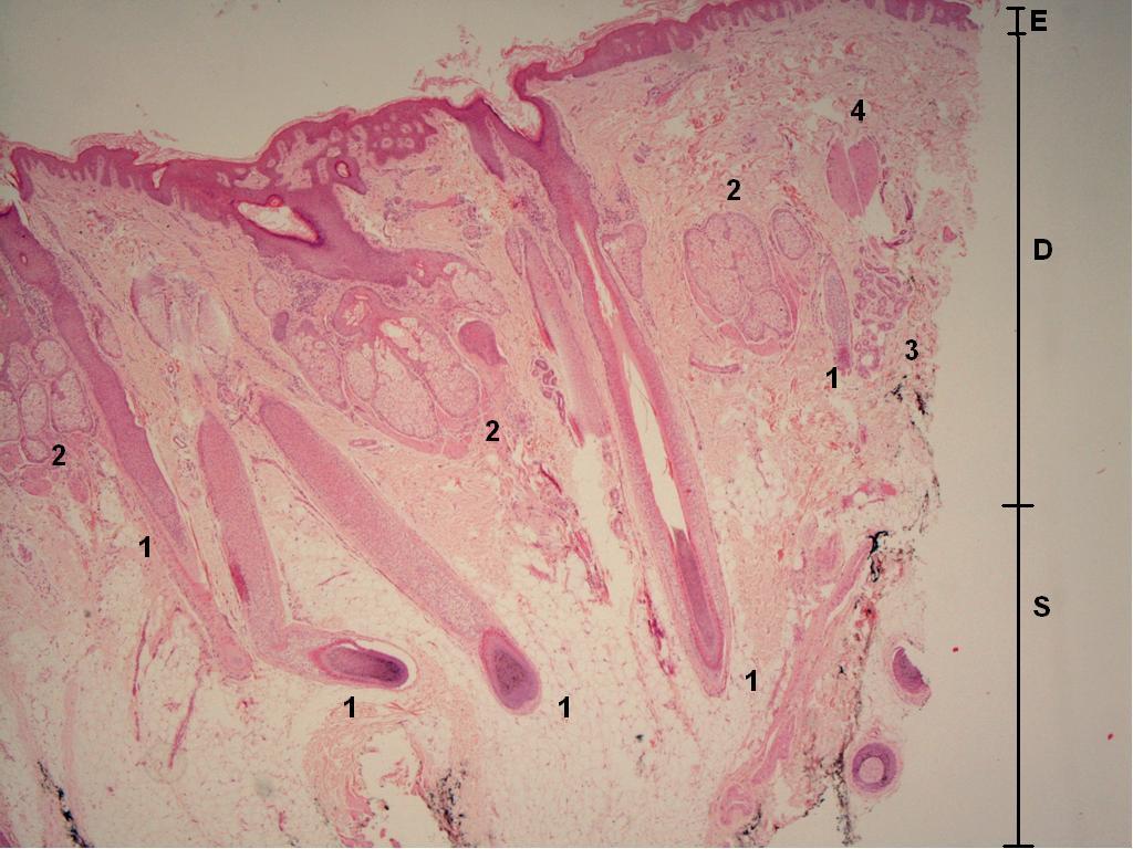
Normal skin
1 - hair follicles,
2 - sebaceous glands,
3 - eccrine glands,
4 - arrector pili;
E - epidermis,
D - dermis,
S - subcutaneous mature adipose tissue
|
|
|
|
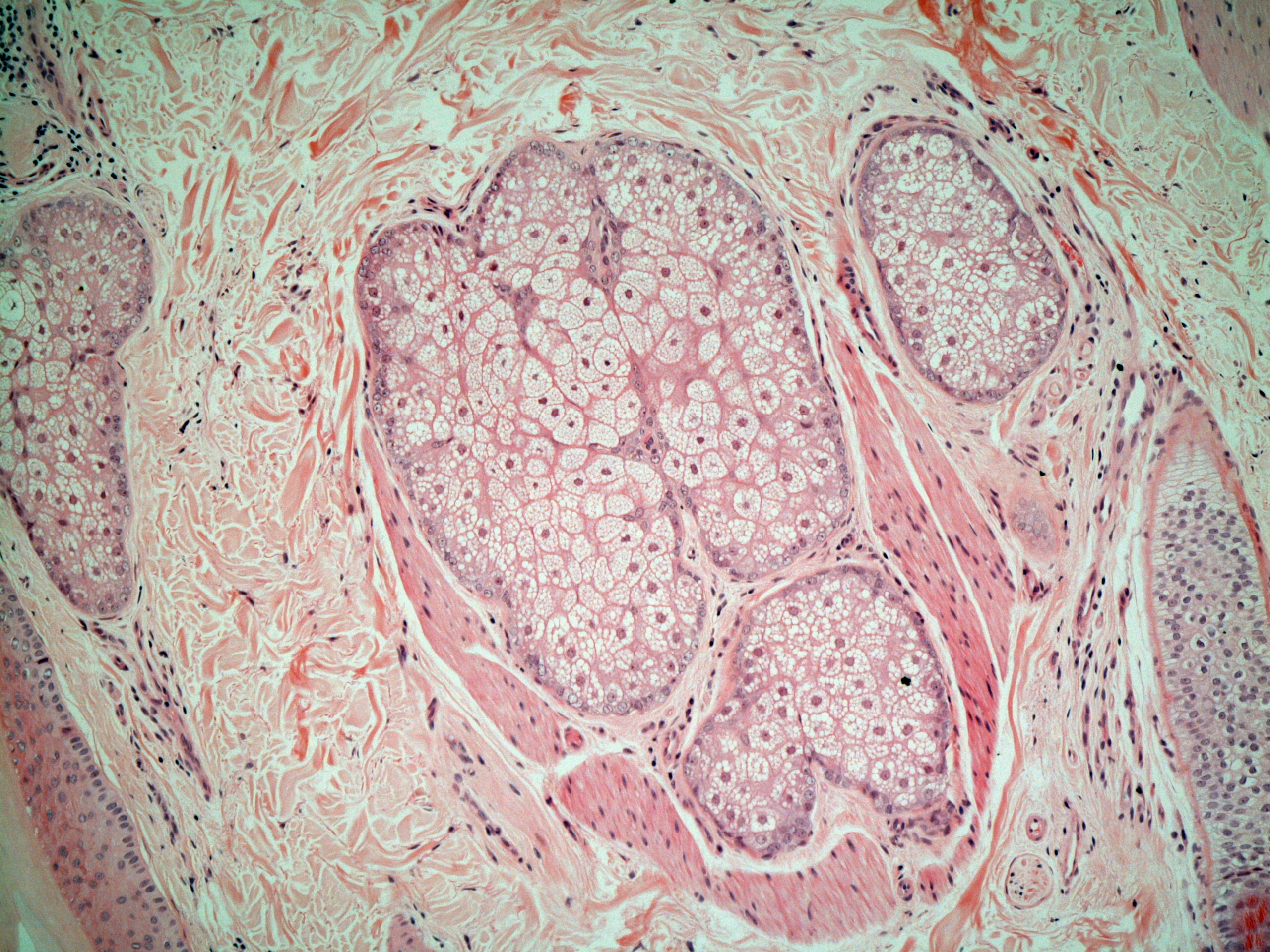
Sebaceous Gland
The cells have vacuolated clear cytoplasm. The outer (germinative) layer of cells is more basaloid and produces the central cells
|
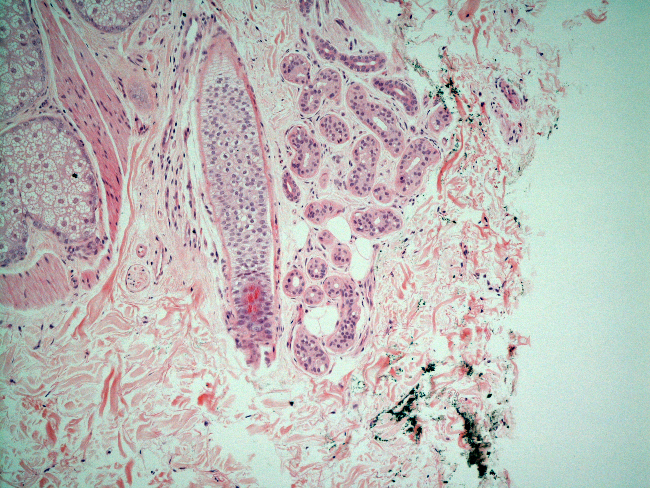
Eccrine Gland
These eccrine glands are situated close to a hair follicle. Their lumens are quite small. At the upper part of the gland, the first part of the eccrine duct is visible. This duct leads to the surface of the skin. In the enlarged image the dual cell layer of the duct can just be appreciated (outer layer - myoepithelial cells, inner layer - columnar cells).
|
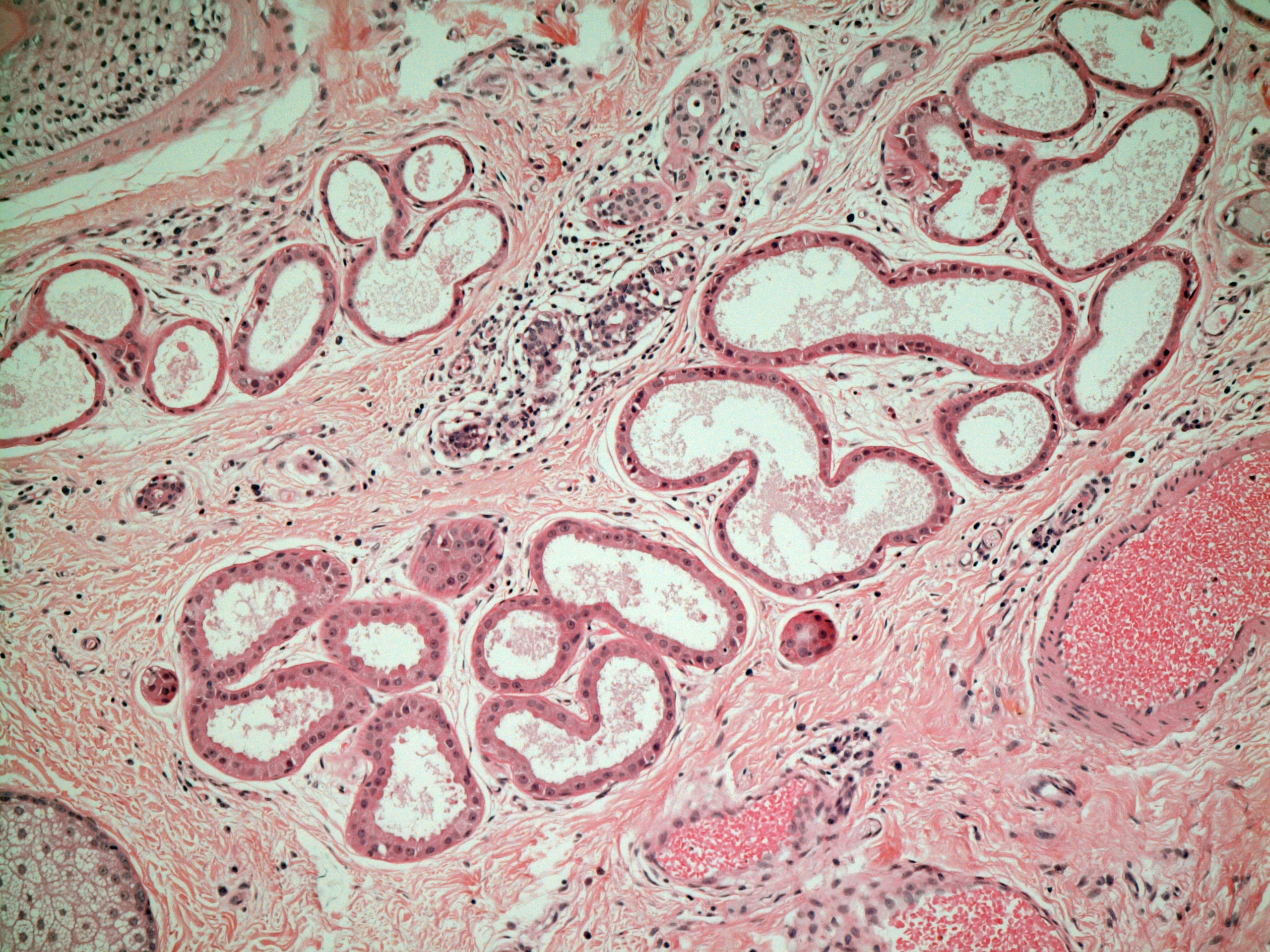
Apocrine Glands
The apocrine glands have wider lumens than the eccrine glands. The cytoplasm of the cells is more eosinophilic. The cells secrete into the lumen by budding off a bleb of cytoplasm at the apex of the cell - decapitation secretion.
|
Physiology
The skin has several functions.
Thermoregulation
The skin forms an insulating barrier to heat loss. Conditions in which there is widespread loss of the skin, such as toxic epidermal necrolysis or burns, can be complicated by excessive heat loss.
The skin also assists in the radiation of excess heat when cooling of the core body temperature is required. Sweating removes heat by the process of evaporation. Shunting of blood from deep vascular networks to superficial plexuses also facilitates radiation of heat. A specialised adapation of blood vessels, known as the glomus apparatus, is involved in this process. The shunting of blood to the superficial vessels explains why a person's skin becomes red when they are exercising and are producing more interal heat through muscle activity.
Fluid Balance
The skin is waterproof and prevents excess loss of water by evaporation from the surface of the body. Widespread loss of skin is often complicated by a marked increase in fluid loss that can be considerably in excess of the basal losses due to normal sweating (around 300ml per day).
Immunity
The skin is part of the innate immune system. Once more this role reflects the skin's ability to act as a barrier. In effect, the skin constitutes a physical obstruction to the entry of bacteria into the body. Breaches in the skin provide a portal of access for bacteria and need to be sealed quickly, hence the initial formation of a scab when the skin suffers a small cut. Scarring is a more dramatic example of the need to seal the breach quickly. A perfect aesthetic result is sacrified in exchange for a more rapid closure of the wound.